

Illumination and microscopy combined in a single fibre bundle by 3D-printed micro-optics
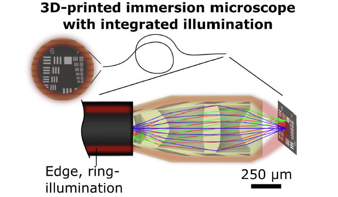
Micro-3D-printed endoscope-tip optics to combine illumination and microscopy in a single device.
GA, UNITED STATES, August 11, 2026 /EINPresswire.com/ -- This article pioneers a new endoscope architecture, reducing the size of ultrathin endoscopes to improve the access to thin luminal organs in endoscopic interventions. Using monolithic micro-3D-printing, a team of researchers integrate a ring-illumination module around a miniaturised multi-lens microscope at the tip of an imaging fiber bundle, with a total device diameter of only ~500 micrometres. The researchers demonstrate the feasibility in lab-scale experiments, and outline the potential of this new endoscopic architecture to produce thinner endoscopes for less invasive future diagnostics.
How can illumination and microscopy work with a single fibre endoscope, just as thin as a particle of ground filter coffee (aeropress/fine pourover grind size)? Using micro-3D-printing, a team of researchers has developed compact fiber-tip optics that co-integrate illumination and imaging in a single fibre bundle endoscope.
Endoscopes usually require separate channels for illumination and bright-field microscopy. For highly miniaturized endoscopes with components in the sub-millimetre regime, the handling and assembly is prohibitively difficult, causing a practical bottleneck for the further miniaturization of endoscopes for biomedical imaging.
This work realizes a micro-3D-printed, monolithic optical system that co-integrates micro-scale imaging and ring-illumination at the tip of an imaging fiber endoscope. Avoiding the difficult manual co-packaging of separate illumination- and imaging channels, this new architecture achieves a compact of just half a millimetre. Additionally, the imaging optics are hermetically sealed, allowing compact imaging with micro-scale resolution both in air and biomedical liquids. Further advances towards practical applicability in biomedical conditions could produce future endoscopes that facilitate the access to small organs and enable less invasive imaging procedures.
References
DOI
10.37188/lam.2026.087
Original Source URL
https://doi.org/10.37188/lam.2026.087
Funding information
This work was supported by the Carl-Zeiss-Stiftung (Nexus 3DEndoFab); the University of Stuttgart (RiSC, Boost Your Science); the Joachim Herz Foundation (Add-on Fellowship) The Baden-Württemberg Stiftung (Elite Programme for Postdocs), Deutsche Forschungsgemeinschaft (DFG, German Research Foundation) – Project number 418911744. Part of this research was conducted at the PETRA III P05 beamline at DESY (Hamburg, Germany), operated by Helmholtz-Zentrum Hereon (beamtime allocated to Proposal 11023209).
Lucy Wang
BioDesign Research
email us here
Legal Disclaimer:
EIN Presswire provides this news content "as is" without warranty of any kind. We do not accept any responsibility or liability for the accuracy, content, images, videos, licenses, completeness, legality, or reliability of the information contained in this article. If you have any complaints or copyright issues related to this article, kindly contact the author above.
